Jan 17, 2023The superficial muscles of the back are responsible for movement of the shoulder. The intermediate muscles of the back assist in the movement of the rib cage during respiration. The intrinsic back muscles facilitate movement of the head and neck and are fundamental in maintaining posture and balance. The posterior or back muscles perform a wide
Fresenius Kabi Introduces Smart Labels for Diprivan® with Embedded Fully Interoperable +RFID Technology | Business Wire
externus. outside. EXternal. internus. inside. INternal. Anatomists name the skeletal muscles according to a number of criteria, each of which describes the muscle in some way. These include naming the muscle after its shape, its size compared to other muscles in the area, its location in the body or the location of its attachments to the

Source Image: ems1.com
Download Image
For the legs, superficial muscles are shown in the anterior view while the posterior view shows both superficial and deep muscles. English labels. From OpenStax book ‘Anatomy and Physiology’, fig. 11.5. Anatomical structures in item: Musculus biceps brachii. Musculus sternocleidomastoideus. Musculus deltoideus. Musculus pectoralis major.

Source Image: edited.com
Download Image
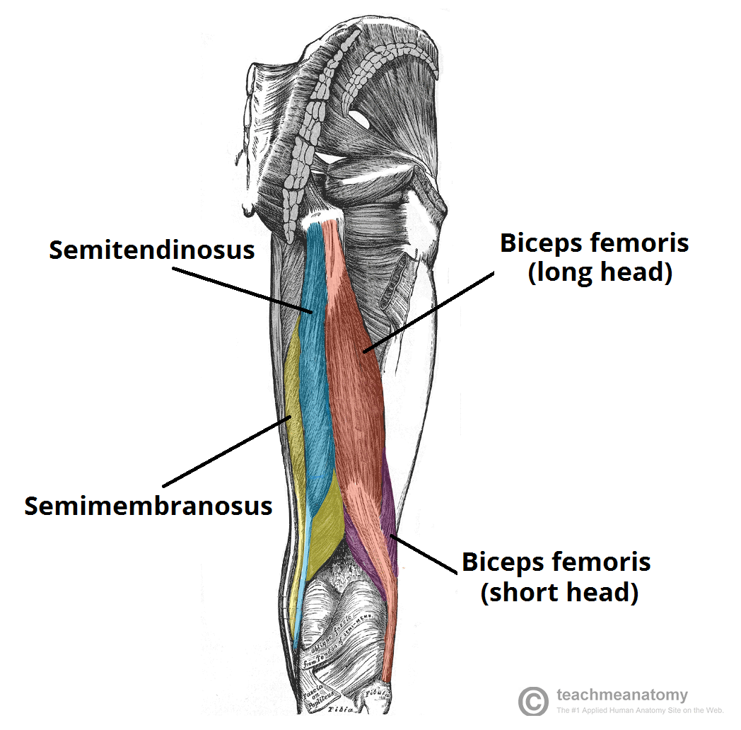
Muscles of the Posterior Thigh – Hamstrings – Damage – TeachMeAnatomy VIDEO ANSWER: We have to show the structures of the shoulder and parlymso in the given figure. The head of numerous, the head of humorous, and the head of the sergical neck are the first living things. Further, further. The scapula is the third one.

Source Image: osmosis.org
Download Image
Correctly Label The Following Muscles Of The Posterior View
VIDEO ANSWER: We have to show the structures of the shoulder and parlymso in the given figure. The head of numerous, the head of humorous, and the head of the sergical neck are the first living things. Further, further. The scapula is the third one. Muscle enabling the hand to extend and to draw near the median axis of the body. Muscle enabling all the fingers, except the thumb, to extend; it also helps the hand to extend on the forearm. Short muscle reinforcing the action of the triceps; it allows the forearm to extend on the arm and also stabilizes the elbow joint. Muscle enabling the
Superficial structures of the neck: Posterior triangle | Osmosis
By the ability of muscle cells to shorten or contract. How do muscle fibres shorten? Sign up and see the remaining cards. It’s free! Start studying Muscles Posterior View, The Muscular System. Learn vocabulary, terms, and more with flashcards, games, and other study tools. 879CBEA2-7E39-4403-BE18-40DF81964E22.jpeg – Correctly label the following muscles of the posterior view . Deep | Superci` Serratus posterior | Course Hero

Source Image: coursehero.com
Download Image
Relaxing your feet affects your hands By the ability of muscle cells to shorten or contract. How do muscle fibres shorten? Sign up and see the remaining cards. It’s free! Start studying Muscles Posterior View, The Muscular System. Learn vocabulary, terms, and more with flashcards, games, and other study tools.

Source Image: frontiersin.org
Download Image
Fresenius Kabi Introduces Smart Labels for Diprivan® with Embedded Fully Interoperable +RFID Technology | Business Wire Jan 17, 2023The superficial muscles of the back are responsible for movement of the shoulder. The intermediate muscles of the back assist in the movement of the rib cage during respiration. The intrinsic back muscles facilitate movement of the head and neck and are fundamental in maintaining posture and balance. The posterior or back muscles perform a wide

Source Image: businesswire.com
Download Image
Muscles of the Posterior Thigh – Hamstrings – Damage – TeachMeAnatomy For the legs, superficial muscles are shown in the anterior view while the posterior view shows both superficial and deep muscles. English labels. From OpenStax book ‘Anatomy and Physiology’, fig. 11.5. Anatomical structures in item: Musculus biceps brachii. Musculus sternocleidomastoideus. Musculus deltoideus. Musculus pectoralis major.

Source Image: teachmeanatomy.info
Download Image
Solved Correctly label the following muscles of the | Chegg.com We are given a picture of a muscle tissue and we are to label the parts of it. Let’s start with the first one. Alright. The first box depends on something. … Correctly label the following muscles of the posterior view: – Flexor hallucis longus – Lateral rotators – Tibialis posterior – Iliotibial band – Serratus anterior – Supraspinatus

Source Image: chegg.com
Download Image
Connect Homework – Chapter 10 Flashcards | Quizlet VIDEO ANSWER: We have to show the structures of the shoulder and parlymso in the given figure. The head of numerous, the head of humorous, and the head of the sergical neck are the first living things. Further, further. The scapula is the third one.

Source Image: quizlet.com
Download Image
Rafael Nadal’s Australian Open withdrawal leaves plenty of questions about his future | AP News Muscle enabling the hand to extend and to draw near the median axis of the body. Muscle enabling all the fingers, except the thumb, to extend; it also helps the hand to extend on the forearm. Short muscle reinforcing the action of the triceps; it allows the forearm to extend on the arm and also stabilizes the elbow joint. Muscle enabling the
Source Image: apnews.com
Download Image
Relaxing your feet affects your hands
Rafael Nadal’s Australian Open withdrawal leaves plenty of questions about his future | AP News externus. outside. EXternal. internus. inside. INternal. Anatomists name the skeletal muscles according to a number of criteria, each of which describes the muscle in some way. These include naming the muscle after its shape, its size compared to other muscles in the area, its location in the body or the location of its attachments to the
Muscles of the Posterior Thigh – Hamstrings – Damage – TeachMeAnatomy Connect Homework – Chapter 10 Flashcards | Quizlet We are given a picture of a muscle tissue and we are to label the parts of it. Let’s start with the first one. Alright. The first box depends on something. … Correctly label the following muscles of the posterior view: – Flexor hallucis longus – Lateral rotators – Tibialis posterior – Iliotibial band – Serratus anterior – Supraspinatus